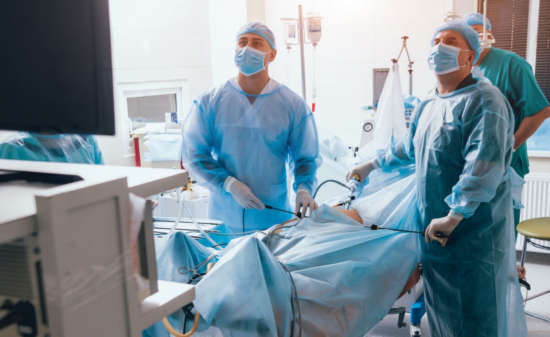
Five small incisions, two of which are 1 centimeter and three of which are half a centimeter, are made on the abdomen. Small cannulas are inserted through these incisions. The abdomen is inflated with carbon dioxide through one of the cannulas and a telescopic camera is inserted and the intra-abdominal image is reflected on the TV monitor. The procedure is performed by entering from other cannulas with surgical instruments. It is a reshaping surgery. It is out of question to cut or remove some of the organs or tissues.
First, the area to be reshaped (lower part of the esophagus and upper stomach) is freed from adjacent tissues.-- In most patients with reflux, the hole through which the esophagus passes through the diaphragm that separates the chest cavity and the abdominal cavity is wide and loose. The angulation in the esophagus-stomach junction was flattened, and the internode in this area shifted upwards towards the chest cavity. This structural deterioration is called sliding hiatal hernia. In the operation, it is ensured that this node is placed under the diaphragm, on the abdominal cavity side, as it should normally be.--The hole in the diaphragm is narrowed with stitches to provide a natural tightness again.(hiatoplasty). --Finally, the swollen, convex upper part of the stomach is wrapped around the lowest part of the esophagus in the form of a collar.(fundoplication) In this way, a high pressure internode (sphincter = gate) mechanism is created again.

